AQUATIC EYES
Underwater vision undergoes some immense evolutionary pressures: turbidity, spectrum of light penetration with depth, temperature, pressure gradients, and the presence of bioluminescence, amongst others (Anca-Narcisa, Ozana-Maria, Cuza et. al 2015). Fittingly, we see a diverse array of eye morphologies underwater, though a common feature amongst all these eyes is that the corneas lie relatively flat – since the refractive index of the surrounding water and the fluid inside the eye are pretty much equivalent, there’s no need for a highly refractive cornea (some aquatic organisms don’t even have one) (2015). Instead, the lens wields the brunt of the focusing power (2015). Eye placement, shape, and size are a variable of the amount of light at a given depth (2015).Surface fish, dolphins, shrimp, and octopi have globose eyes and high-res vision – epipelagic eyes, which have adapted to high levels of exposure, often contain different screening pigments that filter light (Evans, Acosta, Bolstad 2015; see also Frank, Porter 2009).
Meanwhile, in the mesopelagic, we start seeing a reduced visible spectrum of downwelling celestial light. Telescopic eyes start occurring at deeper depths where maximizing exposure of light on the retina becomes more favorable than flexible focusing. Screening pigments are usually absent in these deeper forms, sacrificing resolution for higher sensitivity. Most mesopelagic eyes are rod-cell dominated, as being able to distinguish different visible frequencies is less advantageous in such a dark environment. Whatever cone cells are present are often for viewing the blue light of bioluminescence.
At even deeper depths, the eyes are usually further reduced or nonexistent.

FIG.1– teleost eyes of a yellowfin tuna. Notice the spherical lens, double-layered cornea (outer layer formed by skin), and retractor lentis muscle, which allows for accommodative lens movement (Andison, Sivak 1994). Re-drawn from Walls 1942.
FIG.2– scallop eyes! The “mirror” layer in the eye is made up of highly-reflective guanine crystals that concentrate light into 2 layers of the retina, allowing for complex albeit small-scale imaging (Palmer et. al. 2017). Re-drawn from Speiser et. al. 2011.
FIG.3– crystalline eyes of the mantis shrimp. Re-drawn from Physiologizing 2014.
FIG.4– Sepia cuttlefish eye, which has convergently evolved a shape similar to human eyes (Yoshida et. al 2014). Re-drawn from Ch. 16.1 – Mollusca, ©️The McGraw-Hill Companies, Inc.
FIG.5– Telescopic eye of a hatchetfish Argyropelecus aculeatus, a bathypelagic fish. Re-drawn from Warrant, Lockett 2004.
FIG.6– Amongst the jawless fish, lampreys have surprisingly complex eyes, possessing a 3-layered retina and a melanin-pigmented choroid (Dubielzig 2009). Re-drawn from Dubielzig 2009.
FIG.7– Polychaete worm eyes. Re-drawn from Duke-Elder 1958.
FIG.8– eyes of a mature sturgeonfish; the lens is secured by 2 cartilaginous papillae, but has no observable means of accommodative focus (Dubielzig 2009). Re-drawn from Dubielzig 2009.
Terrestrial eyes are predominantly spherical, though telescopic forms do exist amongst certain nocturnal organisms (2015).
TERRESTRIAL EYES
The first terrestrial ocular systems were presented with unique challenges. On land, the refractive index of the aqueous humor is now greater than that of the surrounding air, meaning that our first amphibious ancestors probably experienced severe nearsightedness when they first clambered to shore. Consequently, selection for strong curvature in the cornea became favorable as it accommodated for refractive differences inside and outside the eye (Anca-Narcisa, Ozana-Maria, Cuza et. al 2015). Now, focusing power becomes an interplay between both cornea and lens (2015).Terrestrial eyes are predominantly spherical, though telescopic forms do exist amongst certain nocturnal organisms (2015).

FIG.1– eye of the oriental garden lizard Calotes versicolor. Re-drawn from Young 1981.
FIG.2– elongated, tubular eye of an owl (upper) as well as a chicken eye (lower) for comparison – despite the difference in form, both eyes contain pecten, a comblike protrusion of blood vessels that maintains pH in the vitreous humor and nourishes the retina – these are a common feature of most bird eyes (Brach 1977). Re-drawn from Martin 2017.
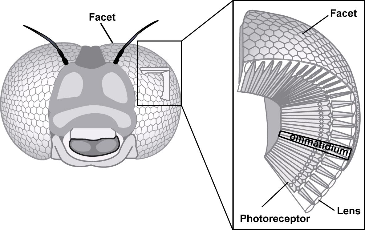
FIG.3– parts of an insect’s compound eye. Re-drawn from Ecole Polytechnique Fédérale de Lausanne.
FIG.4– a frog eye. Re-drawn from Mangold 1931.
FIG.5– a cat’s eye. Re-drawn from Gelatt 2019.
I'll end with some questions I'm planning to look into!
I'll end with some questions I'm planning to look into!
- What's with all the protruding tissue we see in birds and reptiles? Does it hinder light refraction onto the retina in any way?
- Does that ectodermal conus in the reptilian eye have a function? What selective advantages have shaped it this way? (Also, it is a convergent characteristic to pecten, or are the two related somehow...?)
- How many times have fovea evolved independently?
- What's with those certain fish species that can perceive UV light? What evolutionary advantages would this provide underwater?
- What kind of selective pressures drove the evolution of a 3-layered retina in lamprey eyesight? Are there other examples of parasites with good eyes, or is the lamprey a freak exception?
- Why are polychaete eyes shaped like ear endoscopes? And why are sturgeon eyes shaped like sad-looking pastries?
- Eye vs. brain size – positive or negative correlation? Would we get different answers for highly-visual vs. less-visually oriented clades?
- Compound eyes come in a huge variety of patterns, colors, lusters, facet shapes. What environmental factors/niche characteristics influence each quality?
- Determinants of pupil shape?
- When is it more advantageous to have a greater number of eyes rather than eyes that are larger/have better visual acuity and/or range of vision?
SOURCES
Dubielzig, D. (2009). A Survey of Ocular Anatomy and Pathology of Vertebrate Species [PowerPoint slides]. Retrieved from https://www.vetmed.wisc.edu/pbs/dubielzig/pages/coplow/PowerPoints/Wildlife_Dz_Worksh_08.pdf
Warrant, E. Lockett, N. A. (2004). Vision in the Deep Sea. Biological Reviews 79(3):671-712.
Anca-Narcisa, N. Ozana-Maria, P. (2015). “Aquatic” vs. “Terrestrial” Eye Design – A Functional Ecomorphological Approach. Biologie animală, 61: 101-114.
Wilk, L. (2009). Mantis Shrimp Eye Structure and Function, Semantic Scholar. 1-6.
Duke-Elder, S. (1958). System of Opthalmology: The Eye in Evolution. St. Louis, Missouri: C. V. Mosby Company.
Martin, G. R. (2017). What Drives Bird Vision? Bill Control and Predator Detection Overshadow Flight. Frontiers in Neuroscience 1(11):619.
Young, J. Z. (1981). The Life of Vertebrates. Oxford, England: Clarendon.
Gelatt, K. N. (2019). Eye Structure and Function in Cats. Retrieved from https://www.merckvetmanual.com/cat-owners/eye-disorders-of-cats/eye-structure-and-function-in-cats
Fig. 3. Composite eye and ommatidies of an insect [scientific diagram]. From Ecole Polytechnique Fédérale de Lausanne. Retrieved from https://passion-entomologie.fr/wp-content/uploads/2015/12/compound_eye.jpg
Warrant, E. Lockett, N. A. (2004). Vision in the Deep Sea. Biological Reviews 79(3):671-712.
Anca-Narcisa, N. Ozana-Maria, P. (2015). “Aquatic” vs. “Terrestrial” Eye Design – A Functional Ecomorphological Approach. Biologie animală, 61: 101-114.
Wilk, L. (2009). Mantis Shrimp Eye Structure and Function, Semantic Scholar. 1-6.
Duke-Elder, S. (1958). System of Opthalmology: The Eye in Evolution. St. Louis, Missouri: C. V. Mosby Company.
Martin, G. R. (2017). What Drives Bird Vision? Bill Control and Predator Detection Overshadow Flight. Frontiers in Neuroscience 1(11):619.
Young, J. Z. (1981). The Life of Vertebrates. Oxford, England: Clarendon.
Gelatt, K. N. (2019). Eye Structure and Function in Cats. Retrieved from https://www.merckvetmanual.com/cat-owners/eye-disorders-of-cats/eye-structure-and-function-in-cats
Fig. 3. Composite eye and ommatidies of an insect [scientific diagram]. From Ecole Polytechnique Fédérale de Lausanne. Retrieved from https://passion-entomologie.fr/wp-content/uploads/2015/12/compound_eye.jpg
Brach, V. (1977). The Functional Significance of the Avian Pecten: A Review. The Condor 79:321-327.
Palmer, B. A. Taylor, G. J. Brumfeld, V. (2017). The Image-Forming Mirror in the Eye of the Scallop. Science. 358(6367): 1172-1175.
Andison, M. E. Sivak, J. G. (1994). The functional morphology of the retractor lentis muscle of a teleost fish, Astronotus ocellatus. Canadian Journal of Zoology, 1994, 72(11): 1880-1886.
Yoshida, M. Yura, K. Ogura, A. (2014). Cephalopod eye evolution was modulated by the acquisition of Pax-6 splicing variants. Nature, 4 : 4256.
Evans, A. B. Acosta, M. L. (2015). Bolstad, K. S. Retinal Development and Ommin Pigment in the Cranchiid Squid Teuthowenia pellucida (Cephalopoda: Oegopsida). PLOS One, 10(5): 1-11.
Frank, T. Porter, M. (2009). Spectral sensitivity, visual pigments and screening pigments in two life history stages of the ontogenetic migrator Gnathophausia ingens. Journal of the Marine Biological Association of the United Kingdom, 89(1): 119-129.