Thursday, April 30, 2020

Portuguese Man-Of-War

Hi all! The glass jellyfish models caught my attention in John's museum tour because jellyfish are my favorite animals. I decided to research the Portuguese Man-Of-War because I have had more than my share of unfortunate (and painful) encounters with them in the past (many stories to tell... let's just say that although they lack brains, they always find me somehow).

This is what a Portuguese Man-Of-War looks like, and fun fact: it's not technically a jellyfish. The organism is actually siphonophore, a colony of polyps closely related to the jellyfish, and is composed of four different types. These polyps are all genetic clones of one another, and the four types are 1) the ones composing the sail 2) the tentacles (stinging/hunting function) 3) the reproductive organs that are inside of the tentacles and 4) other organisms to carry out metabolic functions

The "sail" on top helps the sea jelly move with the wind, and this first species is what gives the colony its name. Old war ships had sails that sort of resemble this gaseous sac, so this is why the common name is the Portuguese Man-O-War. Although these colonies do not have brains, they can adjust the amount of air inside their floats to submerge.

Reproductive polyps and muscular polyps that pull the food up to the digestive organs are the third and fourth type of polyps. Not as much is known about these, since most scientific queries revolve around the tentacles and gaseous sac for movement.

Due to their bluish color, these creatures are difficult to spot underwater, and the average lengths of their tentacles are around 30 feet! Record lengths have been up to 165 feet. Just because the jelly has washed up on the shore, it is never a good idea to touch it because the nematocysts are probably still active and can still release their venom. The mechanism behind how each barbed stinging cell is the same in any cnidarian: it has a trigger hair at a cellular level (the small tab on the operculum in the image below). These coiled projections are also barbed down the coiled part that springs out, which is a large cause for the skin irritation and tingling pain when being stung.


Each nematocyst is individually triggered, and so if there is enough pressure on a single cell, it will release its barbed projection. This is why dead jellyfish (not just the Portuguese Man-Of-War) can still sting. The charged nematocysts may still be intact and any unfortunate person who touches it or otherwise triggers the cells will receive a painful sting. 

Citations:

https://www.nationalgeographic.com/animals/invertebrates/p/portuguese-man-of-war/


https://oceanservice.noaa.gov/facts/portuguese-man-o-war.html

Citation for cnidarian nematocysts: my high school biology class and 3 years working at the Aquarium of the Pacific.

Research at the Sierra Nevada Aquatic Research Laboratory

First off, I would like to thank John and Claudia for organizing these fun virtual tours, especially the one about the UC reserves. I had always meant to look into the UC reserves and their opportunities, but I would constantly forget to do so. The Bodega Marine Reserve and the Sierra Nevada Aquatic Research Laboratory (SNARL) appealed to me the most. The most interesting to me in terms of undergraduate research, however, is SNARL. I did look into the Bodega Marine Reserve, but I feel like its research in the Non-Indigenous/Invasive Species and Pathogen Facility would be more suitable for grad school.

Researchers at SNARL are currently focusing on the conservation and recovery of mountain yellow-legged frogs, which were previously abundant in the Sierra Nevada. These frog populations are declining due to the fungal disease Chytridiomycosis; the disease’s emergence in the Sierras is attributed to the arrival of invasive species. The team has studied the spread of Bd (the fungi behind Chytrid) through coding, frog re-introductions, infection experiments, immunological studies, and spatial analyses.

This research is interesting to me because it incorporates data about the invasive species who brought Chytrid. Additionally, the mixture of community ecology, disease ecology, and mathematical approaches used in this research attracts me. I would specifically like to learn more about how math is incorporated into this research. Since the reserve is affiliated with UCSB and I have previous experience with Chytrid introduced through invasive species, I am thinking of possibly reaching out to the team for a summer internship or a quarter-long research experience. If this is something of interest to any of you, the link to the official lab page is: https://mountainlakesresearch.com/

Wednesday, April 29, 2020

Missing Organelle: Mitochondria

I'm sure many of us have heard early in our introductory biology classes that "Mitochondria is the powerhouse of the cell". The concept is a simple one, and is often understood by many. However, there are some eukaryotes which have adapted to survive without mitochondria. One such eukaryote Monoceromonoides exilis is similar to most eukaryotic cells, except for its curious missing mitochondria and peroxisomes. This paper seeks to use genomics to examine possible causes and differences between M exilis and other metanomads. 

Interestingly, the proteome of M exilis is believed to be more representative of ancestral metamonads (phylogenetic group). Unlike other metamonads which are characterized by a lack of introns (4-60), M exilis has over 32000 introns in its genome. This has interesting implications as the large number of introns requires greater energy devoted to gene expression. This suggests that despite missing a mitochondria M exilis is able to produce plentiful ATP through substrate level phosphorylation alone.  The paper goes into much more detail than what was listed here, so if any of this interest you I strongly recommend checking it out.

https://academic.oup.com/mbe/article/36/10/2292/5525708#163713785

Mycelium Magic

Mycelium Magic


A few years ago there was a bit of buzz over the potential application of fungi in the growing plastic waste issue, but as with a lot of Facebook post fodder the excitement eventually died down. The products seemed to have potential but there just hadn't been enough research or development of the techniques to apply them right away. 


One question you may ask now is how are things looking several years later


Background:
The call to reduce plastic pollution has been increasing in intensity as study after study shows the tremendous impact single-use plastics can have if they are released into the environment. From microplastics leaching into our natural resources through water and even air pollution to massive garbage patches choking our oceans and marine life, something clearly has to be done. Considering the estimates for the global production of plastics in 2015 were between 380 and 450 million tons, and the fact that these numbers are expected to double by 2050, the transition to more sustainable manufacturing practices is essential. One solution is to fight the issue at its source by encouraging manufacturing companies to make the transition from single-use plastic to more sustainable options. One that may come to mind is shipping material. It can be astounding how much plastic packaging is used just to transport a single product. Even worse, these are generally made from plastics that are extremely difficult to recycle and are certainly not durable enough to be reused by the manufacturer. 


Here’s where the fungi come into play. In 2007 the company Ecovative was started to explore how mycelium can be used to replace these materials. By inoculating agricultural plant waste such as cotton burrs and grain husks with mycelium producing fungi, then heating and drying the material to cease the growth of the mycelium, biodegradable packaging material can be produced at a much lower cost and carbon footprint than that of conventional materials. 


Where are they now


In 2013 Ecovative signed an agreement with Sealed Air Corporation (yes that's the real name) to begin commercializing this manufacturing technique for various consumer and packaging materials. And in 2016 it looked like IKEA would commit to using their packing material. From a quick search on their recent patents, it seems that they have plans for much more than packaging. One other particularly interesting project they have started is to develop plant-based meat alternatives using mycelium. Another update on their website stated they are releasing a cell culture scaffolding that uses mycelium to create 3D tissue culture. This is directed as something for the alternative meat industry, but if this idea actually holds water, this could be extremely useful in the tissue regeneration field as well.


The future of fungi


Judging by the focus of their projects, this company is working on good intentions. By providing a more economical and (better yet) ecological alternative, they can begin to wean our off of plastics. This is a slow-moving process though. IKEA has still yet to begin packaging their furniture with the mycelium alternative. Their ideas are promising but there have still yet to be any major breakthroughs or corporate partnerships to go into production that would bring in the influence they are hoping for. 


While they may have not made any major industrial booms yet, in 2017 they did receive a 9 million dollar US department of defense contract for their materials research. It is important to remember that the amount of time it takes for an idea to finally take root is not a perfect indicator of its merit or viability. And with this in mind, many people still root for Ecovative to make an impact on how we approach the materials we use in our day-to-day life. 


Plastic pollution:


Ecovative:


Tuesday, April 28, 2020

Unicellular Eukaryotes 2: Chimera Boogaloo

In this post I would like to talk about Criptomonad algae, a type of algae that are said to be a mix of two distinct unicellular eukaryotes. The paper I looked at focused a lot on the Endosymbiont Theory of eukaryotic cells that states that, evolutionarily, eukaryotes come from the symbiosis and engulfing of many prokaryotes over time. However, in this paper, the idea is that the Criptomonad algae is made up two unicellular eukaryotes. This discovery was made by using PCR to amplify the Cryptomonas DNA and then adding markers to find similarities to the existing database. Then, to go further, the rRNA was also analyzed and this gave more insight into the evolutionary line and phylogeny of the Criptomonad algae(1). This discovery is important as it supports the endosymbiosis theory, but also shows flaws and misconceptions about the phylogenetic classifications of the Criptomonad's Kingdom, Chromista(1).


1.) Douglas, S., Murphy, C., Spencer, D. et al. Cryptomonad algae are evolutionary chimaeras of two phylogenetically distinct unicellular eukaryotes. Nature 350, 148–151 (1991). https://doi.org/10.1038/350148a0

Monday, April 27, 2020

Crazy Ant Mutualism!!

Hi, y'all! I want to share the paper I found last week, because it was just so cool. It described a new discovery in the complex mutualism between ants and fungi.
     It had long been known that ants cultivate fungus, and the two have a mutualistic relationship. This paper reported that a third member of the relationship had been found: a filamentous bacterium growing on the ants along with the fungus. The bacterium is in the genus Streptomyces. Bacteria in this genus produce the majority of antibiotics currently used for medical purposes in humans. It does the same in this relationship. The bacteria was found to have potent inhibitory effects on Escovopsis, a fungal genus that acts as a specialized parasite of ant fungal gardens. The bacteria grows in the same places on the ants' body, depending on species and type of ant. This suggests that the association is highly evolved, and perhaps ancient in origin. The fact that the bacteria is transmitted vertically (from parents to offspring) and promotes growth of the fungal mutualist in vitro also suggests that this mutualism has been around for a very long time.
     This paper is from 1999, so it's pretty old, but the relationship described is absolutely fascinating to me. I hope y'all think so too! Have a good week five!


Bioassay of Streptomyces inhibition of Escovopsis

Source: Currie, Cameron R., Scott, James A., Summerbell, Richard C., Malloch, David. Fungus-growing ants use antibiotic-producing bacteria to control garden parasites. (1999). Nature 398: 701-704. 

Diatoms in Forensics

Hi all! This week I was looking into diatoms because I had never really researched them that much, and John had briefly mentioned them under the unicellular eukaryotes umbrella.  I found many cool articles on how diatoms are used in forensics, and how they can serve as legal proof to aid in determining the cause of death for a person.

When a body is found in a body of water, it can be tested for the presence of diatoms within the organs. If diatoms are found within a body, then it is likely that the person drowned. The diatoms would have entered the body while the person still has respiratory activity, and it would be more difficult for the diatoms to enter the body post-mortem (Lunetta "Autopsy", Verma).  Diatoms are easily traceable marine organisms, due to them being microscopic, and their silicon structures resist decomposition long after the body has started to decay (Verma). According to Lunetta, the species of diatom found within the body can also help narrow down the areas of water where the person may have drowned ("Autopsy").

However, it is possible to have a false positive, since after soft tissues of the body decay, it is possible for diatoms to become present in bone marrow from merely being exposed to the body long enough (Lunetta "False-Positive"). Verma also notes that the absence of diatoms does not automatically rule out drowning as a cause of death, either, since people with heart or lung conditions, or who hyperventilate, are less likely to inhale water before drowning. 

This may have been a more morbid subject for the week, but I had no idea that diatoms were used  for forensics, and this just really stuck out to me as a cool way to use non-human species as clues to solve a puzzle.


Lunetta, P. “Autopsy Findings: Drowning and Submersion Deaths.” Encyclopedia of Forensic and Legal Medicine, 2016, pp. 315–322., doi:10.1016/b978-0-12-800034-2.00048-3.
Lunetta, Philippe, et al. “False-Positive Diatom Test: A Real Challenge? A Post-Mortem Study Using Standardized Protocols.” Legal Medicine, vol. 15, no. 5, 2013, pp. 229–234., doi:10.1016/j.legalmed.2013.03.002.
Verma, Kapil. “Role of Diatoms in the World of Forensic Science.” Journal of Forensic Research, vol. 04, no. 02, 2013, doi:10.4172/2157-7145.1000181.

My Amazing Experience at Landels-Hill Big Creek Reserve!

Hi everyone! This blog post is a bit different than my others that normally contain some cool research I've found while looking into one of the weekly topics; however, this week, I was able to look into the UCNRS and reminisce in my time spent in Big Sur!

In Summer 2018 right after my freshman year at UCSB, I had the opportunity to take part in one of the Wildlands Studies field programs in Big Sur. I spent two weeks backpacking and camping around Big Creek Reserve while conducting a Steelhead trout survey, a sea otter population census, several plant transects, and learning about the different species of intertidal organisms.

For this post, I looked back at my field notebook from this research and thought I'd share some reflections from day 1 of field surveys in streams:
"The trends in Steelhead populations vary a lot depending on depths of pools, different coverings of plants, and different ground coverings."
"Stream field work was much more difficult physically than I was expecting because the pools we were analyzing were not clearly marked, and the cliffs we had to climb down to get to them were steep and riddled with poison oak. Nicole said that because we were walking up and down in our 8mm wetsuits, running shoes, and full face masks, that we would be protected from the poison oak - but it was definitely extremely tiring."
"Our methods included one person [we did stream surveys in groups of two] drawing a map of the pool while the other counted the fish; then, the second person would count the fish based on size and estimated age. We then took sediment percentages together, and measured the pools to make accurate maps later with transect tape."
"Today we say a massive animal skeleton at the bottom of one of our pools. Salomé [my partner] discovered it and I heard screaming from her snorkel. It looked huge with our masks, and we were kind of grossed out because we had been sitting in the same pool of water as this decomposing organism with fish eating it."

The work I did at Big Creek Reserve was definitely some of the hardest physical conditions I've worked in, but it was definitely worth it! I constantly look back at my time here and remember how much I learned in what feels like so little time. I definitely look forward to doing more field work in the future and hope to return to Big Creek some day soon! If you have any questions about Wildlands feel free to reach out:)

Southern ocean phytoplankton: is there hope?


This week in the video on microbial eukaryotes, we talked about phytoplankton and their limitations of growth. The biggest limiting factor was said to be the low iron levels in oceans. One use of iron in phytoplankton is in the synthesis of chlorophyll. In the polar regions, phytoplankton also have a limiting factor of low temperature and low light. What's worse is that to adapt to low light conditions, it would seem like a good way would be to have an increased iron intake, to produce more chlorophyll for harvesting light. It seems that any southern ocean phytoplankton that dare to try to survive and thrive would be met with this twisted positive feedback loop. All in all, it seems that southern ocean phytoplankton really seem to have the odds stacked up against them...

But, when the (selection) pressure is high, there are some species that just manage to adapt and succeed!

I found a paper published in 2019, showing that some phytoplankton have actually managed to adapt to these harsh conditions. The paper reports up to a six-fold increase in photosynthetic rate. What's more interesting is the method by which they did this. The paper found that they had one of the largest photosynthetic antennae recorded for a phytoplankton species. Under normal low light conditions, having a large photosynthetic complex is usually disadvantageous. However, coupled with the low temperature of the southern ocean, it seems that this disadvantage is mitigated and phytoplankton are able to survive!

Works cited

Strzepek, R. F., Boyd, P. W., & Sunda, W. G. (2019). Photosynthetic adaptation to low iron, light and temperature in Southern Ocean phytoplankton. Proceedings of the National Academy of Sciences, 116(10), 4388-4393. doi:10.1073/pnas.1810886116

Sunday, April 26, 2020

Pink (Red) Snow

Hello everyone!

You may recall the momentous news that I shared with you this week- snow can taste like watermelon! Unfortunately, I regret to inform you that despite searching the internet for a least 20 minutes, I could find absolutely no scientific evidence that pink snow does taste like watermelon. :(

However, I did find an article that stated in a laboratory trial of 7 healthy individuals aged 24-56 who voluntarily ingested 500g of pink snow experienced no significant episodes of gastrointestinal distress (Fiore, McKee, and Janiga 1997)! Is this the 100% go signal for you to try consuming pink snow on your own to see if you think it tastes like watermelon? Eh, maybe not because there's plenty of other things lurking in the snow you may not really want to ingest, but ultimately the choice is yours. If you do choose to try it out, please let me know- I would really like my experience to be corroborated...

In any case, during my deep dive into the intricacies of red/pink snow, I found out a lot of interesting information! Red snow is actually a species of green algae- Chlamydomonas nivalis which utilizes high concentrations of carotenoid pigments to protect it from the UV rays at high elevations. A great informative and quick read regarding Chlamydomonas and related species is Milius (2000).

Finally, before I let you go thinking "Oh yeah, red snow- great stuff!" I must also inform you of the significance of red snow and the shrinking arctic ice. Lutz et al. (2016) showed the correlation between high concentrations of red algae and bacterial communities and a decreasing albedo of snow and ice in the arctic. The decreasing albedo in the arctic is one of the primary causes of higher annual melting rates, so studying the snow microbiome will become increasingly important in the context of global warming and climate change. If you want to get a better understanding of the breakdown of the microbial community of the arctic, I would highly recommend you check out this article.


References;
Fiore, David C., Denise D. McKee, and Mark A. Janiga. 1997. “Red Snow: Is It Safe to Eat? A Pilot Study.” Wilderness & Environmental Medicine 8 (2): 94–95. https://doi.org/10.1580/1080-6032(1997)008[0094:RSIIST]2.3.CO;2.
Lutz, Stefanie, Alexandre M. Anesio, Rob Raiswell, Arwyn Edwards, Rob J. Newton, Fiona Gill, and Liane G. Benning. 2016. “The Biogeography of Red Snow Microbiomes and Their Role in Melting Arctic Glaciers.” Nature Communications 7 (1): 11968. https://doi.org/10.1038/ncomms11968.
Milius, Susan. 2000. “Red Snow, Green Snow.” Science News 157 (21): 328. https://doi.org/10.2307/4012510.


Zombie Ants and Their Fungal Overlords

This week I looked into cordyceps and their crazy ability to colonize and control their insect hosts. I was first introduced to cordyceps as a little kid through a nature documentary (probably narrated by David Attenborough as all the best ones are), and I actually saw one in person in Costa Rica! It had taken over a leafcutter ant was glued to a leaf by the fungus and the ant's own bite.

The genus cordyceps was created to define the group of fungal pathogens of arthropods, characterized by the ascomata or stromata that rise out of the insect cadaver. I looked into the species Ophiocordyceps unilateralis, which specifically targets ants from the tribe Camponotini. I thought this species was particularly interesting because I'd seen something like it before and I found out that this one is able to exist inside the host for a while without any symptoms, and then can dramatically alter the ant host's behavior before it dies.  Once Ophiocordyceps unilateralis makes its way into the host's body, there is an incubation period in which all seems normal. This allows the fungus to thwart the social immunity strategy of ant colonies. Normally, sick ants are cast out for the good of the colony, but Ophiocordyceps unilateralis is undetectable until it is too late. After the incubation period, the fungus takes hold of the ant's nervous system and controls its muscles through bioactive compounds. It forces the ant to move to a humid microclimate and then bite into whatever plant it is on and wait for death. Ophiocordyceps unilateralis feeds on the cadaver and then extends its fruiting body through the head of the ant. If this isn't dramatic enough for y'all, I'll link a very suspenseful and dramatic video by National Geographic detailing this process.

Getting to the bottom of the zombie ant phenomenon | Penn State ...

Cordyceps Are Cool: Here's a video to prove it

The life cycle of Bd, the fungus associated with the global amphibian decline.

This week I read Life cycle stages of the amphibian chytrid Batrachochytrium dendrobatidis. Bd is one of major topics of study of the Briggs and since I'm now a part of it, I thought it'd be useful and interesting to read about the life cycle...and it's a fungus! I'd highly recommend checking out the photos of the fungus in the article.

Bd is part of the Chytridiomycota division of fungi. It causes an epidermal infection of fungi, and it's the only member of its phylum to cause disease in vertebrates. The life cycle is relatively simple. The zoospore, which is the flagellated stage, grows into a thallus, which then produces a zoosporangium, which releases more zoospores. Bd is not known to reproduce sexually. In an amphibian the immature zoosporangia start in the epidermal cells in the lower layers and zoosporangia mature with the epidermal cell as it moves to the upper layers. They complete their development in the dead keratinized cell on the surface. The discharge tubes develop towards the skin surface, and release the spores into the environment.

The study tried to answer why the fungus has the capacity to kill frogs when it's restricted to the epidermis, and why it is less susceptible to topical antifungals when in amphibian skin. They did ultrastructural studies, basically looking at the fungus in skin under the microscope, to find out why.

They describe the life cycle in more detail: The zoospore disperses, it resorbs its flagellum and develops into a germling, which has fine rhizoids which branch from it. This develops into the thallus stage, which is the stationary stage that produces the zoospores.  This is crazy: Over time there are mitotic devisions making the the thallus multinucleate, and then there is cleavage and the thallus becomes full of flagellated zoospores. The swollen part of the thallus with the zoospores is the zoosporangium. A discharge tube also develops and the zoospores escape after a plug dissolves. Empty zoosporangia remain. Sometimes zoospores that did not escape grow in the zoosporangia, and sometimes bacteria enter and replicate. They also say that thalli grow better together, and zoospores alone usually die. In the epidermis up to 3 sporangia were in a single epidermal cell. Sometimes mature sporangia don't make it past cornified cell layers, and end up discharging zoospores into intercellular spaces. Often Bd is found in clusters on the skin too.

Bd infections caused roughening of the skin, and often there were bacteria in between cells and in discharged sporangia. Infected frogs had uneven layers of skin and epidermal cell maturation seemed disrupted. There was also a build up of keratinized cells that would normally be shed. Sporangia also displaced the nucleus of infected cells and changed fibril structure in cytoplasm. The sporangia seemed to cause the premature death of the cells. These effects could be the reason for the lower susceptibility of  Bd to antifungals in the epidermis.

They did not determine what actually causes Bd to kill its host but clearly they cause some unhealthy skin.
This article was written in 2005, so I did a quick google search to see if someone had discovered the reason. This website states that Bd can inhibit electrolyte transport which can lead to cardiac arrest

Citation:
Berger L, Hyatt A, Spear R, Longcore J. (2005). Life cycle stages of the amphibian chytrid Batrachochytrium dendrobatidis. Diseases of Aquatic Organisms. Vol. 68: 51–63
DOI: 10.3354/dao068051

Friday, April 24, 2020

Satellite remote sensing of phytoplankton

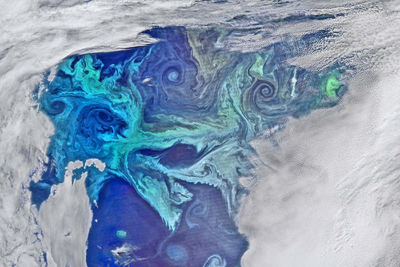
An important eukaryote, phytoplankton not only support the marine food web and fisheries, but also play a big role in the Earth’s carbon cycle, acting as a carbon dioxide sink and producing half of the oxygen we need to breathe. Phytoplankton’s far-reaching importance makes it crucial for researchers to observe the dynamics of their population abundance and distribution. One method of studying these single celled eukaryotes is through using satellite remote sensing. Radiance data from satellites measure chlorophyll (Chl) pigment concentration which is a good representation of variance in marine primary productivity, causing it to be an indicator of phytoplankton biomass. 
Until recently, satellite data indicated general phytoplankton concentration but could not be utilized to distinguish between different types of phytoplankton. Researchers from the Alfred Wegener Institute Helmholtz Centre for Polar and Marine Research developed an algorithm that can be used to categorize the phytoplankton from the satellite imagery into five groups. These groups were determined by analyzing how the amount of sunlight that hits the ocean and is reflected back into space (i.e. reflectance) depends on the type of phytoplankton. By detecting variations in reflectance, the algorithm can be used to fingerprint the type of phytoplankton present in any marine region. It must be noted that direct water samples are still required to verify the species. 
Another way that satellite data can be used is to look at how disturbances, such as coastal upwelling, can imbalance phytoplankton’s tight predator-prey relationships and result in phytoplankton blooms. Data from NASA’s satellite, MODIS, was used to display how places with upwelling have a greater phytoplankton abundance. The environmental conditions that phytoplankton rely on, including upwelling, are impacted by climate change. However, satellite-derived phytoplankton concentration records span back to 1979, making it difficult to observe long-term trends. In the recent Nature paper, “Global phytoplankton decline over the past century,” Daniel Boyce et al. describe how in situ Chl measurements since 1899 suggest a link between increased sea surface temperatures and decreased Chl trends. Advancements in phytoplankton research methods, such as satellite remote sensing, and further studies on the relationships between phytoplankton and environmental conditions are necessary for a better understanding of macroecological changes in the ocean. 
If anyone is interested in getting their hands on some of this satellite data, let me know because I found some cool resources while looking through the literature. Also, check out NASA’s worldview map with Chl data from their MODIS satellite.


Sources


Alfred Wegener Institute, Helmholtz Centre for Polar and Marine Research. "Observing phytoplankton via satellite." ScienceDaily. 19 March 2020. www.sciencedaily.com/releases/2020/03/200319125151.htm.

Boyce, D., Lewis, M. & Worm, B. Global phytoplankton decline over the past century. Nature 466, 591–596 (2010). https://doi.org/10.1038/nature09268

Garner, Rob. “NASA Ocean Data Shows 'Climate Dance' of Plankton.” NASA, 29 Sept. 2014, www.nasa.gov/content/goddard/nasa-ocean-data-shows-climate-dance-of-plankton/.

Monday, April 20, 2020

SOFeX: The Iron Fertilization Experiment


For this week’s topic, I was super interested in the Southern Ocean Iron Enrichment Experiment (SOFeX) done by principal investigators from the Moss Landing Marine Laboratory and the Monterey Bay Aquarium Research Institute in 2002. This experiment was designed to test the Martin Hypothesis, which stated that an increase in iron dust in the ocean would cause phytoplankton blooms since iron is a limiting resource in their growth. Their increasing biomass could then sequester CO2 out of the atmosphere, cooling the earth and combating climate change. The reason they performed this experiment in the Southern Ocean was because those waters were high in nitrate, which isn’t a limiting resource for algal growth, yet low chlorophyll (or phytoplankton biomass), indicating that they’re most likely iron-limited. They performed this experiment in two different locations, one north that low silicate concentration and one south that has high. Silicate is used by diatoms to make shells, so it is also a potential limiting resource that would denounce the Iron hypothesis. There had been 2 previous iron experiments done before SOFeX, but this experiment wanted to determine the phytoplankton’s capacity to significantly impact the carbon cycle. In order for iron to have a positive effect on CO2 sequestration, the phytoplankton need to be eaten and travel to the deep sea, instead of just being decomposed and letting all the CO2 go again. 

Now for the results: the experiment found massive blooms in the photosynthetic community due to the influx of the growth limiting resource. The phytoplankton productivity was dominated by nonsilicate plankton in the north vs. diatoms in the south, which is explained by the different levels of silicic acid in the waters. Both biomasses exported CO2 from the atmosphere with higher iron input, also proving that iron played the most dominant role in limiting growth, and showed that iron inputs could significantly impact the atmospheric carbon cycle. 


Citations:
Kenneth H. Coale, et al. Southern ocean iron enrichment experiment: carbon cycling in high-and low-Si waters. Science. Vol. 304 #5669 (April 16, 2004).
“SOFeX Cruise History & Purpose.” MBARI, Monterey Bay Aquarium Research Institute, www3.mbari.org/expeditions/SOFeX2002/history&purpose.htm.

Sunday, April 19, 2020

Parasites that make hosts Zombies

Have you ever wondered what it would be like if zombies existed in real life? Well, there are definitely some parasites that make this happen. These parasites are the ones in charge of modifying the behavior of their hosts and controlling their actions. These change of behaviors vary from organism to organism and can range from making the host go to its predator or behaviors that benefit the host.

According to the article, parasites change their hosts' personalities, which in turn, their behavior is altered. But,  how are these interactions modified? The article references a study in which they came to the conclusion that this control is made by the parasite's ability to control neurochemical pathways as well as its immune responses.

One example of these organisms are the insect parasitoids. These parasites exploit their hosts by making the hosts become "bodyguards." As a consequence, the hosts behave in a way in which they protect and feed larvae. This helps the parasites, as they can use the infected hosts as pawns to protect the parasites. One such parasite is the jewel wasp, that can inject lethal chemicals that paralyze cockroaches and then proceeds to inject venom to the cockroaches brains in a precise manner to control its ability to walk. All while the wasp lays its eggs inside the cockroach for incubation.

Embedded Image

Another really interesting example is the Toxoplasma parasite. This parasite is known to infect mice, cats and other animals. The shocking part is that some cases of Schizophrenia in humans are caused by this parasite. As this parasite also make mice develop Schizophrenia, many scientists believe mice to be a good model organism to further study the effects of this parasite as well as Schizophrenia in general. Mice affected by this parasite also lose fear of cats! Furthermore, it is thought that as these infected mice are eaten by cats, the cats also become infected. How do humans become infected? It is hypothesized that feces of infected cats are the primary source of spread of this parasite to humans!

Sources:
Knight, Kathryn. "How pernicious parasites turn victims into zombies." (2013): i-iv.

Lichenicolous Fungi

Hey, y'all! Hope everyone had an okay week 3, despite everything being extremely wacky right now.
     This week, I took an interest in lichens. The symbiosis between algae, fungi, and bacteria was really interesting to me. I looked into lichenicolous fungi, which are fungi that form obligate associations with lichens.
     Lichenicolous fungi are most often either parasites of lichens, or sapotrophs that colonize decaying lichen matter. Something really interesting to me is their host specificity. The paper I read said that "as many as 95% [of lichenicolous fungi are] thought to be associated with only a single lichen genus" (Lawrey and Diederich). Additionally, some groups of lichen have more host-specific lichenicolous fungi associated with them than others. Certain groups may make for better hosts because of their chemical or nutritional properties, their wide geographical distribution, or even their relatively higher availability in evolutionary history. This last factor would seem to suggest that more ancient species of lichens would have a larger number of host-specific lichenicolous fungi.
     The paper also discusses the ecological effects of lichenicolous fungi's high host specificity. Lichen parasites with high host specificity are not a threat to species of lichens other than their host, so they have very specific effects. Lichen groups with a large number of host-specific parasites could go two ways. The first, which lines up with the theory of a coevolutionary origin, is that many lichen species will have evolved, having fewer, but very well-adapted, parasite species. The second, which lines up with the independent adaptation theory, is that the group's lichens will have a wider variety of parasites.
     Host specificity is what interested me the most about this paper, but it talks about a lot of other topics related to the biodiversity and evolution of lichenicolous fungi. If this subject interests you even a little, this paper is a fantastic place to start!

image source: lichenicolousfungi.net 

Source: Lawrey, James D., Diederich, Paul. Lichenicolous Fungi: Interactions, Evolution, and Biodiversity. (2003). The Bryologist 106(1): 80-120. 
   

Saturday, April 18, 2020

Intraspecific Competition Between Different Strains of Trypanosoma brucei brucei


Intraspecific and interspecific competition between parasites is one of my research interests, partly because of my strange obsession with parasites (my family is tired of me talking about them during dinner) and how experimental evidence on competition between them is scarce. So, when parasites and the two types of competition were mentioned during this week’s lectures, I immediately knew what I wanted to focus on. I decided to look into one of my favorite parasites, Trypanosoma brucei, which causes African Sleeping Sickness in humans and other animals (usually livestock). This paper focused on two strains of Trypanosoma brucei brucei, which were isolated from different hosts. One strain (later dyed green) was gathered from a hartebeest while the second (later dyed red) strain was gathered from a tsetse fly. For those of you interested, I attached a photo above so you can see how cool the parasites look!
 
To keep this short, the researchers found that intraspecific variation in multiple strains can alter the behavior of the parasite population and its effects on the host, demonstrating that multiple-strain infections are vastly different from single-strain infections. Intraspecific interactions between these strains could lead to changes in host-population dynamics. Mutual suppression of these strains can also affect transmission rates because it changes the concentration of parasite strains a vector consumes. According to the authors, this mutual suppression of strains also alleviates the parasite’s effects on the host. However, these parasite strains may evolve to out-compete each other by increasing their virulence. There’s a lot to this paper and I mainly wanted to summarize some key points, so I’ll attach a citation below if you want to check out the specific methods and findings. :)

Citation:
Balmer, O., Stearns, S.C., Schötzau, A. and Brun, R. (2009), Intraspecific competition between co‐infecting parasite strains enhances host survival in African trypanosomes. Ecology, 90: 3367-3378. doi:10.1890/08-2291.1

Friday, April 17, 2020

The "Mimic Octopus"

Remember that huge school of fish in Finding Nemo that make shapes like the Sydney Opera House and other animals and whatnot? Pretty cool, and also pretty far-fetched. Or so I thought.
I present, the so-called "mimic octopus." It has such immense phenotypic plasticity that, beyond camouflage, it can actually mimic the behaviors of other organisms in its environment. Though the actual mimic octopus is, as its name implies, Thaumoctopus mimicus, many types of octopus have been observed performing mimicry behaviors, such as Macrotritopus defilippi in the Atlantic and Octopus cyanea in the Indo-Pacific. These creatures have been videotaped mimicking organisms like flounders, parrotfishes, sea snakes, and rays. Of course, which organisms they choose to mimic is dependent upon their given environment and their present needs. While the mimic octopus was mentioned in the textbook as an example of predation-driven adaptations, I think that it's also quite a cool example of neurobiology, in that switching between different phenotypes within a split second illustrates very precise neural control. As explained by Hanlon, Forsythe, and Joneschild in their 1999 paper (cited below), the speed at which certain octopuses are able to transition between extremely specific behavioral phenotypes implies a straightforward and fast-acting neural circuit. This mimicry behavior of really emphasizes the connections between gene expression, phenotype/behavior, and community interactions, which I think is quite amazing. There's something radical to be explored at every level of biology regarding these mimic octopuses (it seems that papers call them octopuses and not octopi). Also, this behavior is super cool to watch, so check it out below!

https://www.youtube.com/watch?v=Wos8kouz810

Mimic octopus mimicking a sea krate, a lionfish, and flounder ...


Hanlon, R.T., Forsythe, J.W., & Joneschild, D.E. (1999). Crypsis, conspicuousness, mimicry and polyphenism as antipredator defences of foraging octopuses on Indo-Pacific coral reefs, with a method of quantifying crypsis from video tapes. Biological Journal of the Linnean Society, 66(1), 1-22.

Hanlon, R. T., Watson, A. C., & Barbosa, A. (2010). A “mimic octopus” in the Atlantic: flatfish mimicry and camouflage by Macrotritopus defilippi. The Biological Bulletin, 218(1), 15-24.

Wednesday, April 15, 2020

Mating of Peacock Spiders

     When Claudia was talking about new species, I found the peacock spider particularly interesting. I would probably be super scared of this thing, but it definitely looks pretty cool from a safe distance:). I found an article that discussed peacock spider mating rituals, which like the peacock bird, are particularly elaborate. An observational study done at UCBerkeley looked at mating trials under different vibratory and lighting conditions. Ultimately, what they ended up finding was that female mate choices are not impacted by longer wavelengths nor vibration signals.
     Lots of animals use mating signals that humans may not even notice, such as polarization and UV light signals. Typically, male peacock spiders uses colorful flaps on the abdomen and waves these flaps along with legs at the targeted female, as well as using vibrations in song. Comparing peacock spiders to other jumping spiders, this visual display and vibratory movements are pretty common among other jumping spiders. In this study, which measured male color and irradiance factors through 175 mating trials (86 successful), one factor that seemed to play a large role in mating success was overall brightness levels. Furthermore, the vibration sounds were found to complement the visual signals and were sometimes useful in mating rituals.
     One major takeaway from this study as well as John and Claudia's lectures seems to be that behavioral ecology is extremely complex and scientists have barely scratched the surface in understanding lots of animal behavior. This peacock spider example is one of many that indicates the complexity and expansiveness of courtship display. One direction the researchers of this study will likely look into are response signals from female peacock spiders in terms of decision making. I look forward to seeing where this data will take them, as I am now more curious than ever about visual display in mating ritual.
Here is a figure from the journal that shows how they looked at pigmentation of the spider to analyze vibrance data.

Girard, MB., Kasumovic, MM., Elias, DO. (2018). The role of red coloration and song in peacock spider courtship: Insights into complex signaling systems. Behavioral Ecology, 29(6), 1234-1244. http://dx.doi.org/10.1093/beheco/ary128

Monday, April 13, 2020

On the Uncertainty of Summer

Ok gang,
For a while I had no idea what I wanted to do for my blog post, but after a
conversation with friends about concerns over how summer research will
be affected by the news of the online summer instruction, I wanted to try and
give some hope or encouragement


To reiterate what I said to the chat:
“... science and art aren’t things to be suspended because of a lack of
money or gathering place, it will persist in whatever form it can, for however
long it has to”


To better explain what I mean by that, here are some major historical
contributions made under isolation, quarantine, or times of disease:

- Shakespeare wrote many sonnets during a plague outbreak around
1592, and possibly King Lear and Macbeth when the playhouses shut down
for a total of 78 months due to plague outbreaks between1603 and 1613

- A young Isaac Newton developed theories on optics, prisms, and began his work on papers that would become the foundation to calculus while in isolation due to the plague sending everyone home from university
- Edvard Munch not only contracted Spanish Flu but also began making self portraits of his appearance in recovery as soon as he was well enough to hold a brush
  • This could be anything from working with existing data that can be
used for a different guiding question to anything within your field that 
           doesn't require your boots on the ground
for this advice are the people you work with, reach out!
- Lit Review some of you may have done this in high school but now’s the time
to make it fun
  • Try and find papers that interest you and are in your reach of skill (or
          not that's fine too)
  • Think of how they may mesh together and could possibly point
towards a question you can work on from home or prepare for the fall
walls of your room will judge your ambition
whatever seems counterintuitive, and then look into why it makes
you feel that way, you may be surprised with what you find
- Online Coding course
  • Code academy: Pro is giving some free trials for students 
  • DataCamp: first chapter is free
  • PyCharm: not quite a lesson plan, but a very powerful tool for playing 
          around with python once you get a bit of a base in it
  • ACCESS THE UCSB VPN! Especially if your lab needs you to have
cnsi account


This isn't to say that COVID-19 should make you the next legendary scientist,
playwright, or artist, but it does tell you that our lives, both academic and
otherwise, are far from put to a halt. Do what you want: shave your head, crochet
a tree, scream obscenities at the water running from the faucet, the world is
your oyster just make sure you don’t cook it. It is easy to fall into a feeling of
lacking control, and a pet project may be just what you need. Whatever that
may be to you.


If you're wondering what you can do in terms of "research" or any other remote
biology work:  
Here's what little I have to offer. If anyone has other things to add please
comment them as they would be welcome additions. Even if its just interesting
youtube channels, books, or podcasts.

- If you are already in a lab and have yet to hear from them, reach out to
your mentor about work you can do remotely 
    • we all inhabit a very wide range of subjects, so the best people to ask
    • Don't be afraid do get interdisciplinary with it, it's not like the cold blank
    • look at the textbook and focus on what doesn’t make sense
      • Not what you can't quite grasp (though that’s important too), but


    In the end even if you are not doing "research" you can still be building yourself
    as a researcher (or whatever it is you feel like being) in isolation. You don't need
    a grant or lab bench to do the work you love.

    Also:


    The best choice for you may be to just ignore everything I just said and do
    what you want

    Go for it!
    • Take what advice will help you, leave the rest
    • Look to the others around you and within yourself
    • Do whatever seems right for you


    Hope everyone is doing well


    Link for Campus VPN


    FREE Code Academy PRO COVID 19 link