In class, we learned about a mechanism that plants have in order to sense light around them. The proteins in chatge of this are phytochromes these respond to light. As explained in class, these proteins respond to far red and red light. This is interesting from an engineering point of view because we can engineer these proteins as delivery vehicles. This technique is called optogenetics and is the main technique used in my laboratory. The following image shows how the proteins PhyB and PIF can be used to cluster proteins of interest (POI) using light:
What I thought it was really interesting was the fact that these proteins that react with light not only exist in eukaryotes but prokaryotes as well. One example is that bacteria use visible light in order to activate mechanisms of DNA repair. These different proteins are "activated" with different colors of light. For instance, the phytochromes discussed earlier react with red and far red light
whereas others such as the protein "Cry2" react with blue light and deactivate in the dark. My personal project uses one of these proteins that activate with blue light in order to control the behavior of a set of proteins, called "SMAD," by allowing them to reach the nucleus and activate transctiption. We also can make them do the opposite behavior. The proteins that allow me to control such behaviors with light are really important for both the engineering and biology fields as they allow me control individual cells with light and are reversible (meaning that they can be turned on or off as I wish).
Sources:
1. De Mena, Lorena,
Patrick Rizk, and Diego E. Rincon-Limas. "Bringing
light to transcription: the optogenetics repertoire." Frontiers in
genetics 9 (2018): 518.
2. Repina, Nicole A., et al. "Engineered illumination devices for optogenetic control of cellular signaling dynamics." bioRxiv (2019): 675892.
3. OptoBase
For Biology students in the College of Creative Studies at the University of California Santa Barbara.
Tuesday, March 17, 2020
Mutant Baby Jellyfish
Hey y'all, Marina can't log in so this is her blog post.


In my lab last week I was tasked with inducing strobilation in Aurelia aurita polyps, and while I was sorting out the baby jellyfish (their proper name is ephyra but I call them jebbies too) I noticed a bunch of mutants, Hell Yeah!!
They seem to come in 3 common types:
This is a plate full of normal ephyra for reference:
- 4 appendages instead of 8
- 12 appendages instead of 8
- ?????
Type A and B seem to be of a similar type of mutation. Taking a stab at why exactly these mutations were to occur, one may start out with hypothesising that whatever genetic mechanism determining appendage number may require two copies in order to produce the wild type. This could mean that type A may be caused by only having 1 copy while type B is caused by having 3. I have no idea if this is true as I was unable to find much online in terms of publications, this is just the quiet speculation of an undergrad.
Phototactic Symbionts in Coral
Hey y'all: Marina can't log in, so this is one of her blog posts.
You may know already that coral have a lot of active fluorescent proteins which is already pretty cool, but did you know they can act as beacons to attract the Symbiodinium that live and photosynthesise within them?
Symbiodinium are dinoflagellates that live within the coral polyp and provide ~80% of the nutrients required by the coral. With this fact in mind you may think these Symbiodinium are teeming in the water on reefs but they are actually quite low in concentration. The question is then raised in how polyps acquire these microbiota that are so essential for survival.
I’ll drop the link below for this paper I was sent that examines just that question. Researchers were able to demonstrate the both positive and negative phototactic tendencies of Symbiodinium and how those can assist the coral in attracting them. In this paper they mainly focused on the attraction to green fluorescent protein which is very common in most coral. But in reference to my other post on the Glowing Glowing Gone campaign, the phototactic tendencies may play a role in why these coral may start to fluoresce when they bleach (lose their Symbiodinium) What should be remembered though is that the Symbiodinium studied in this paper and negatively phototactic to blue and purple so for this hunch to pan out there would have to be more attention paid to a wider range of Symbiodinium.
Glowing Glowing Gone
Hey y'all: Marina can't log in, so this is one of her blog posts.

Preface: If you have not seen the Netflix documentary Chasing Coral please do!! It’s beautifully shot/edited and the approach to bring awareness to the coral bleaching epidemic is extremely compelling
Having seen the documentary ~6 times so far, what never ceases to get me is the section shot right off the coast of New Caledonia. While on the mission of documenting the coral bleaching event of 2016 on the Great Barrier Reef, the team was tipped off about a region in which the already bleached coral developed strangely vibrant and intense fluorescent tones. Off the cuff, the team studying the bleaching hypothesised that this could be a last ditch effort by the coral to protect itself from the increasingly damaging UV rays and heat that was causing the bleaching in the first place. (whoa)
Inspired by these vibrant colors, the creators behind the documentary began the design campaign “Glowing Glowing Gone” to bring awareness to the increasingly concerning bleaching events occurring on coral reefs. The campaign challenges designers and artists to use the three main tones of fluorescence exhibited by these bleaching coral in their pieces. I highly recommend checking out the account on instagram and the link to their website below.
Sunday, March 15, 2020
Wash your hands

With fear of coronavirus spreading like a pandemic, governments, companies, and school campuses are implementing aggressive campaigns to curb the spread of the virus. Although social distancing and avoiding large gatherings are usually mentioned, these campaigns seem to universally focus on handwashing as the key way to stop the virus. Why is washing your hands so important, and why does it kill coronavirus?
![]() |
| The structure of a virus |
As it turns out, viruses that are nearly impossible to destroy once they've infected you are quite easily to kill when they're exposed. Viruses are pretty simple. Nucleic acids (single stranded RNA for coronavirus) and a few proteins necessary for infection are surrounded by a lipid bilayer. Transmembrane proteins involved in infection cover the membrane. When you wash with soap and water, soap (an amphiphiles) has polar ends that interact with water and nonpolar ends that interact with the lipid bilayer. This causes the membrane to dissolve and the virus is inactivated. Wash your hands!
![]() |
| Transmission EM of coronavirus. Glycoproteins poke out from the virus' membrane. |
sources:
https://bionewscentral.com/researchers-trace-coronavirus-outbreak-in-china-to-snakes/
https://www.phta.org/news/coronavirus
https://www.theguardian.com/commentisfree/2020/mar/12/science-soap-kills-coronavirus-alcohol-based-disinfectants
Campbell biology 11th edition
Saturday, March 14, 2020
Mutualistic Relationship between sloth hair and bacterial community
I’m writing about the mutualistic relationship between cyanobacteria and eukaryotes in sloth hair and sloths. In my research, I discovered that sloth hair is not just home to various types of cyanobacteria, but also diatoms, fungi, macro-organisms, and algae. Specifically, the green algae Trichophilus welckeri is the most common algae found in the fur of three-toed sloths (Suutari et al.). The algae is believed to provide a green discoloration of the fur that allows them to camouflage with the forest canopy. According to Suutari et al., “among all 71 sloth individuals studied, 73% hosted algae in their fur,” supporting the fact that the algae is frequent and abundant on sloth fur (Suutari et al.). This is most likely due to the fact that three-toed sloth hair has cracks that allow the algae to grow and colonize in the presence of rainwater absorbed by the hairs (Pauli). It is also speculated that the sloth might receive nutrients through diffusion and absorption through the algae, and allows for even more beneficial bacteria to grow (Suutari et al.). Data shows that, since the alga has not been found in many juveniles, the acquisition of T. welckeri happens when the mother passes it to her offspring through physical contact and grooming (Suutari et al.).
Not only does sloth fur have a symbiotic relationship with alga, it also supports a big fungal microbiome, much bigger than the human body supports. Higginbotham et al. studied this microbiome in the hopes of finding medicinal qualities to investigate and use practically. The study found through isolating many strains of fungi that “many of these isolates display bioactivity in vitro against parasites that cause malaria and Chagas disease, breast cancer cells, and both Gram-positive and Gram-negative human pathogenic bacteria,” (Higginbotham et al.). The fur of these sloths that live in close proximity are also home to diverse, bioactive fungi that play a cryptic role in maintaining the ecosystem that thrives in sloth hairs.
The study by Pauli et al. about mutualism in sloths presents interesting data on a three-way mutualistic relationship between three-toed sloths, pyralid moths, and T. welckeri. It observed the dangerous and cost-ineffective behavior of sloths descending from their trees to defecate in the ground once a week, and hypothesized that there must be a mutualism that rewards the sloth for that behavior. Indeed, they found that sloth fur was a mating ground for pyralid moths, and that when they would defecate, the moths lay their larvae in their excrement. The moths in the fur also acted as a link between the sloth and its environment, providing it with vital nutrients and increasing nitrogen levels in the fur, which allowed the algal communities to grow as well. Pauli et al. found that “microalgae were three to five times richer in lipid content,” and that “a food item with this high lipid composition would provide an especially rich [...] and rapid source of energy to sloths,” who have a nutritionally poor diet that leads to low energy and metabolism levels (Pauli). This way, the sloths acted as a way for moths to reproduce, and in return they offered nutrients to algae in the sloth’s fur, which the sloth would eat to supplement their diet, developing a three-way mutualism.Citations:
Suutari, M., Majaneva, M., Fewer, D.P. et al. Molecular evidence for a diverse green algal community growing in the hair of sloths and a specific association with Trichophilus welckeri(Chlorophyta, Ulvophyceae). BMC Evol Biol 10, 86 (2010). https://doi.org/10.1186/1471-2148-10-86
Pauli JN, Mendoza JE, Steffan SA, Carey CC, Weimer PJ, Peery MZ. 2014 A syndrome of mutualism reinforces the lifestyle of a sloth. Proc. R. Soc. B 281: 20133006. http://dx.doi.org/10.1098/rspb.2013.3006
Higginbotham S, Wong WR, Linington RG, Spadafora C, Iturrado L, et al. (2014) Sloth Hair as a Novel Source of Fungi with Potent Anti-Parasitic, Anti-Cancer and Anti-Bacterial Bioactivity. PLOS ONE 9(1): e84549. https://doi.org/10.1371/journal.pone.0084549
Cryo-EM microscopy revolutionizing protein structure characterization
For decades, x-ray crystallography has been the standard for determining molecular structures, including the structure of DNA (see below). X-ray crystallography requires that the target be highly concentrated, and then transformed into a stable crystal structure. By bombarding the crystal with X-rays, and mathematically analyzing the diffraction pattern, it's possible to determine the structure of the molecule.
X-ray crystallography was at the heart of many biological discoveries, but falls short in certain situations. For example, the targeted protein has to be able to form a stable crystal, and there's no guarantee that structure of the protein while in that crystal is the same as its in vivo conformation. For many proteins, it's difficult or impossible to obtain highly concentrated samples of the protein. X-ray crystallography works well for small, stable structures.
Cryogenic electron microscopy (cryo-EM works best where X-ray crystallography falls short, on dilute samples of large proteins. In cryo-EM, a wire mesh is coated in the solution with the protein, which is then dipped in liquid ethane to quickly freeze it in place. Next, a stream of electrons passes through the protein, and the diffraction pattern of the electrons is determined. Where the electrons passed straight through, there is an empty space in the protein. In spots where the electron is blocked or significantly deflected, atoms exist. With hundreds or thousands of these images, computer programs can recreate the 3D structure of the protein with high precision.
The mathematics behind converting thousands of electron micrographs to 3D structures are intensive and quite difficult (especially for a biologist), which presents problems for reproducing results and makes cryo em somewhat inaccessible. Because the mathematics are so difficult, scientists have to make choices on how they transform the data, which makes it prone to systematic error and conformation bias. Additionally, more experienced users are able to generate far more accurate and precise 3D structures than less experienced scientists.
sources:
https://www.nature.com/articles/d41586-018-06791-6
https://www.nature.com/articles/nature12822
https://science.sciencemag.org/content/358/6363/eaao0464/tab-pdf
![]() |
| Rosiland Franklin's famous "photo 51," an x-ray crystallography pattern that revealed the double helix structure of DNA |
X-ray crystallography was at the heart of many biological discoveries, but falls short in certain situations. For example, the targeted protein has to be able to form a stable crystal, and there's no guarantee that structure of the protein while in that crystal is the same as its in vivo conformation. For many proteins, it's difficult or impossible to obtain highly concentrated samples of the protein. X-ray crystallography works well for small, stable structures.
Cryogenic electron microscopy (cryo-EM works best where X-ray crystallography falls short, on dilute samples of large proteins. In cryo-EM, a wire mesh is coated in the solution with the protein, which is then dipped in liquid ethane to quickly freeze it in place. Next, a stream of electrons passes through the protein, and the diffraction pattern of the electrons is determined. Where the electrons passed straight through, there is an empty space in the protein. In spots where the electron is blocked or significantly deflected, atoms exist. With hundreds or thousands of these images, computer programs can recreate the 3D structure of the protein with high precision.
![]() |
| Raw electron diffraction pattern from cryo EM |
![]() |
| 3D structure of a protein derived from cryo EM |
sources:
https://www.nature.com/articles/d41586-018-06791-6
https://www.nature.com/articles/nature12822
https://science.sciencemag.org/content/358/6363/eaao0464/tab-pdf
Friday, March 13, 2020
The Importance of Natural History Collections
After our class trip to CCBER, and my own work in the collections of CCBER and the Santa Barbara Museum of Natural History, I wanted to write about the importance of maintaining collections. The CCBER collection was previously unused and stored in a basement on campus, prior to its discovery and subsequent revamping in 2015.
As scientists looking to use data in our research, we need to have access to well-kept data. As data moves towards being online and easily accessible, less thought (and funding) goes towards the physical collected specimens. However, these collections are extremely valuable for use.
Technological advancements allow for more to be done with specimens, and for more data to be gathered from specimens. In the past, collected bee specimen would simply represent the presence of a species in an area, or the interaction between one species of bee and one species of plant. Today, advancements in pollen DNA metabarcoding are allowing researchers to collect pollen samples from old (already curated) bee specimens and analyze all of the plant species that bee had been visiting. Endangered bees can be analyzed to see how we can change our current ecosystems to better support them. Old specimens can be used to help determine the presence of plant species in an area.
Through this technique, our simple collected data of "bee in location" can be expanded into broader, potentially more useful data. If we abandon the maintenance of collections, we will limit our ability to utilize future advancements in technology. And collections need to be actively maintained, because they can easily be destroyed by dermestid beetles (the family of beetles that taxidermists use to strip the flesh from bones!).
Source:
Behm, R., & Seltmann, K. (2017). From rejection to collection! A new entomology collection at the University of California, Santa Barbara Museum of Natural History takes the university by swarm. UC Santa Barbara: Cheadle Center for Biodiversity and Ecological Restoration. Retrieved from https://escholarship.org/uc/item/90j9s4jh
As scientists looking to use data in our research, we need to have access to well-kept data. As data moves towards being online and easily accessible, less thought (and funding) goes towards the physical collected specimens. However, these collections are extremely valuable for use.
Technological advancements allow for more to be done with specimens, and for more data to be gathered from specimens. In the past, collected bee specimen would simply represent the presence of a species in an area, or the interaction between one species of bee and one species of plant. Today, advancements in pollen DNA metabarcoding are allowing researchers to collect pollen samples from old (already curated) bee specimens and analyze all of the plant species that bee had been visiting. Endangered bees can be analyzed to see how we can change our current ecosystems to better support them. Old specimens can be used to help determine the presence of plant species in an area.
Through this technique, our simple collected data of "bee in location" can be expanded into broader, potentially more useful data. If we abandon the maintenance of collections, we will limit our ability to utilize future advancements in technology. And collections need to be actively maintained, because they can easily be destroyed by dermestid beetles (the family of beetles that taxidermists use to strip the flesh from bones!).
Source:
Behm, R., & Seltmann, K. (2017). From rejection to collection! A new entomology collection at the University of California, Santa Barbara Museum of Natural History takes the university by swarm. UC Santa Barbara: Cheadle Center for Biodiversity and Ecological Restoration. Retrieved from https://escholarship.org/uc/item/90j9s4jh
Ancient Bristlecone Pines as Historians
One of the topics that I found interesting from week 8 was the field of dendrochronology, using tree rings to get data on past events and environmental change. The Ancient Bristlecone Pine Forest in California has been used to gather important data. Though the oldest living trees are over 4,000 years old, we can construct a record that dates further back by using pieces of dead trees and essentially lining up the growth rings that overlap between them. This way, our time period of data extends back about 10,000 years.
The dendrochronological dating methods have even complemented radiocarbon dating. By trying to radioactively date old trees, for which we already know the accurate age since death via cross-referencing the growth rings, scientists were able to calibrate the carbon dating process, and get more accurate results. Because of this, some previously radioactively dated specimens were updated with new ages, and these old trees helped "rewrite history."
Sources:
Ancient Bristlecone Pine Natural History. (n.d.). Retrieved March 13, 2020, from https://www.fs.usda.gov/wps/portal/fsinternet/cs/ (compressed link)
Ancient Bristlecone Pine Forest. (n.d.). Retrieved March 13, 2020, from https://www.fs.usda.gov/wps/portal/fsinternet/ (compressed link)
The dendrochronological dating methods have even complemented radiocarbon dating. By trying to radioactively date old trees, for which we already know the accurate age since death via cross-referencing the growth rings, scientists were able to calibrate the carbon dating process, and get more accurate results. Because of this, some previously radioactively dated specimens were updated with new ages, and these old trees helped "rewrite history."
Sources:
Ancient Bristlecone Pine Natural History. (n.d.). Retrieved March 13, 2020, from https://www.fs.usda.gov/wps/portal/fsinternet/cs/ (compressed link)
Ancient Bristlecone Pine Forest. (n.d.). Retrieved March 13, 2020, from https://www.fs.usda.gov/wps/portal/fsinternet/ (compressed link)
Thursday, March 12, 2020
Permian Extinction
Hey everyone!
I hope everyone is doing well with all the chaos going on recently. As a distraction, I thought I'd write a little about the Permian extinction from a paper I read. For those of you who don't know the Permian extinction was a mass extinction event that in the last 600 million years. We saw a loss of 95% of Earth's population at the time. There are many explanations of what caused the Permian extinction, but the two most popular are volcanic eruptions or an asteroid. The most likely explanation is a volcanic eruption leading to the Permian extinction. We believe that during the time of the Permian extinction, global warming caused the Earth's atmosphere to increase by 6 degrees Celsius and a huge amount of light carbon in the atmosphere. Scientists believe this caused a worsening feedback loop which exacerbated global warming.
There are currently 4 important conclusions made towards the Permian period and extinction.
1. The Permian and Triassic (PTr) boundary is estimated to be around 251 million years ago.
2. The Siberian traps, large vats of volcanic lava, is predicted to have reached peak eruption sometime near the PTr boundary.
3. By examining rocks, scientists have been able to find patterns of environmental changes throughout the late Permian and Triassic periods.
4. Studies of stable isotopes, such as Carbon and Oxygen, reveal a common story of environmental turmoil.
If any of this interests you I strongly suggest you check out the paper!
https://www.sciencedirect.com/science/article/pii/S0169534703000934
I hope everyone is doing well with all the chaos going on recently. As a distraction, I thought I'd write a little about the Permian extinction from a paper I read. For those of you who don't know the Permian extinction was a mass extinction event that in the last 600 million years. We saw a loss of 95% of Earth's population at the time. There are many explanations of what caused the Permian extinction, but the two most popular are volcanic eruptions or an asteroid. The most likely explanation is a volcanic eruption leading to the Permian extinction. We believe that during the time of the Permian extinction, global warming caused the Earth's atmosphere to increase by 6 degrees Celsius and a huge amount of light carbon in the atmosphere. Scientists believe this caused a worsening feedback loop which exacerbated global warming.
There are currently 4 important conclusions made towards the Permian period and extinction.
1. The Permian and Triassic (PTr) boundary is estimated to be around 251 million years ago.
2. The Siberian traps, large vats of volcanic lava, is predicted to have reached peak eruption sometime near the PTr boundary.
3. By examining rocks, scientists have been able to find patterns of environmental changes throughout the late Permian and Triassic periods.
4. Studies of stable isotopes, such as Carbon and Oxygen, reveal a common story of environmental turmoil.
If any of this interests you I strongly suggest you check out the paper!
https://www.sciencedirect.com/science/article/pii/S0169534703000934
Benton, M., & Twitchett, R. (2003, May 07). How to kill (almost all) life: The end-Permian extinction event. Retrieved March 11, 2020, from https://sciencedirect.com/science/articlle/pii/S0169534703000934
Wednesday, March 11, 2020
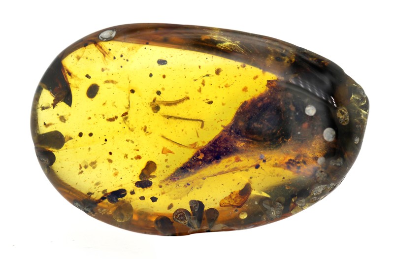
In the news: This miniature skull belonged to a 2-gram dinosaur
I found this in Nature's newest publication and I thought it was interesting! And reasonably fitting too since we just learned about macro evolution.
Preserved in amber, this small bird was found to be 100 million years old, placing it in the center of the cretaceous period on earth. During a time when evolution generally favoured the largest animals and plants to ever grace our planet, it's amazing that a lineage seemed to evolve in the exact opposite direction! However it should be stated that paleontologists admit that there is a bias toward the finding of larger fossils, as they are easier to find. Which makes this an even more incredible find
This small bird weighed in at around 2 grams and was only 2 centimetres long! What's even more fun is that the bird was incredibly well preserved, meaning that it can be subject to analysis and can give amazing data that can help to further contextualize this period of time. Morphological analysis has shown that it contains features not previously observed in any bird.
Due to the bias of finding larger fossils, there are likely many phylogenetic groups that haven't been explored yet, so it's always interesting when we find new ones.
References
Xing, L., O'Connor, J.K., Schmitz, L. et al. Hummingbird-sized dinosaur from the Cretaceous period of Myanmar. Nature 579, 245-249 (2020). https://doi.org/10.1038/s41586-020-2068-4
When to run and when to tumble: bacterial chemotaxis
You're a bacterium, and need to find food, avoid an toxin, or seek out oxygen. Without a nose, brain, and ears, how do you decide which way to go? It's well established that bacteria use a "run and tumble" approach to finding directions. When the bacterium is moving toward an attractant or away from a repellent, it continues in a straight line. However, if the bacterium begins to go in the "wrong" direction, it tumbles (moves turns in a random direction) and tries again.
![]() |
| Run and tumble bacterial chemotaxis. Webre et. al 2003 |
But how do bacteria tell whether they like the direction they're moving in? The ingenious process by which bacteria sense the rate of change in the concentration of chemicals is an astounding example of evolution selecting streamlined and effective strategies in prokaryotes.
![]() |
| Bacterial chemotaxis. Wadhams et. al 2004 |
Ligands bind to MCPs (methyl-accepting chemotaxis proteins) on the cell membrane. When the concentration of an attractant decreases, more MCPs enter their unbound conformation. In this state, MCPs can bind to CheA. CheA is a phosphatase, but is activated by unbound MCPs. So a reduction in ligand concentration increases the amount of active CheA. CheA has two phosphorylation targets: CheY and CheB. when CheA phosphorylates CheY, CheY-P binds to the flagella and reverses its direction of motion, causing the bacterium to 'tumble.' CheZ, which is always present and active, gradually desphosphorylates CheY so that the tumble phase doesn't last forever. After a couple seconds, CheZ has dephosphorylated enough CheY to return the bacterium to its 'run' state. From an evolutionary perspective, the problem is that this can only measure some threshold in the amount of ligand, rather than a change in the amount of ligand. There must be a way to tell MCP that the new level of ligand is 'normal' and that the system should measure deviations from it. The key is that CheA also phosphorylates CheB, which demethylates MCP and makes it more sensitive to ligands.
When the concentration of a ligand increases, the reverse happens. CheA doesn't bind to MCP, so CheY is never phosphorylated and the motor direction never flips. Another protein, CheR, is constantly methylating MCP (desensitizing it to ligand binding), such that the concentration of protein must be constantly increasing to prevent CheA binding.
With this system, there's evidence that bacteria can detect a difference of a single molecule binding or detaching from a receptor, even when the overall concentration of the ligand is very high in the solution.
Wadhams, G., Armitage, J. Making sense of it all: bacterial chemotaxis. Nat Rev Mol Cell Biol 5, 1024–1037 (2004). https://doi.org/10.1038/nrm1524
Seasonal Affective Disorder (SAD)
Hi everyone,
Fitting acronym for how this quarter's been so far....but I'm also interested in psychology and found this neat.
Seasonal Affective Disorder, abbreviated as SAD, is generally marked as yearly depressive episodes falling under winter/late fall, and improving during spring and summer. Originally dismissed as "wintertime blues," SAD has proved to become a mental health problem that affects more people than expected. Interestingly enough, light therapy is cited as one of the best treatments for it, but other options such as cognitive-behavioral therapy, exercise, and medications are available.
SAD seems to increase with latitude in North America, but this trend doesn't match with other countries around the world. It is believed that in the winter, serotonin turnover is lowest, contributing to decreased mood during the months; experiments conducted with light therapy and serotonin have shown improvement, and other separate ones have indicated a correlation between bright lights and improvement in therapy.
It would be interesting to see if other countries around the world have similar rates of SAD in comparison to America, but not much extensive research about this topic has been conducted.
Good luck with finals everyone!
Fitting acronym for how this quarter's been so far....but I'm also interested in psychology and found this neat.
Seasonal Affective Disorder, abbreviated as SAD, is generally marked as yearly depressive episodes falling under winter/late fall, and improving during spring and summer. Originally dismissed as "wintertime blues," SAD has proved to become a mental health problem that affects more people than expected. Interestingly enough, light therapy is cited as one of the best treatments for it, but other options such as cognitive-behavioral therapy, exercise, and medications are available.
SAD seems to increase with latitude in North America, but this trend doesn't match with other countries around the world. It is believed that in the winter, serotonin turnover is lowest, contributing to decreased mood during the months; experiments conducted with light therapy and serotonin have shown improvement, and other separate ones have indicated a correlation between bright lights and improvement in therapy.
It would be interesting to see if other countries around the world have similar rates of SAD in comparison to America, but not much extensive research about this topic has been conducted.
Good luck with finals everyone!
In regards to uncertainty about spring break and next quarter
Hi everyone, I know this isn't quite a regular post, but alas we are in an odd period of limbo and uncertainty. I'm not sure how many of you (especially the first years, but theoretically this could apply to anybody in university housing/whose home is out of state) might be facing uncertainty about your living situation over Spring Break/during the start of next quarter, so I figure I'd extend an offer to anyone who might need it. My home is in NorCal, and we have an extra bedroom. If you need a place to stay for any reason (i.e. canceled/adjusted travel plans so you can stay in-state) please send me an email or if you see me in person wave me down. I happen to work in one of the Chi-5 and I know there's a bit of uncertainty in the housing department right now about what will happen for the start of next quarter (for the time being people will be allowed back, but it's always good to have a backup plan), so if there's any way I can help out I feel obligated to. My email is megrim719@gmail.com so please reach out to me if there's anything I can do for you!
Anyways, here's a cute cat cuz it's all gonna be okay:
https://gfycat.com/forcefuldemandingbarracuda-cats-of-instagram-adorable-kawaii
Anyways, here's a cute cat cuz it's all gonna be okay:
https://gfycat.com/forcefuldemandingbarracuda-cats-of-instagram-adorable-kawaii
Tuesday, March 10, 2020
Purple Non-Sulfur Bacteria Enrichment
For this post, I will be talking about another one of my enrichments and selective processes that I learned about in my EEMB150 lab. For this post, my focus will be on my enrichment for Purple Non-Sulfur Oxidizing bacteria, or PNSBs. Samples of mud were taken from the lagoon on campus and put into two tubes; one with sodium succinate and one with sodium benzoate, both with ammonium for nitrogen sourcing. These two solutions are carbon-rich and both are organic carbons. These bacteria are anaerobic photoheterotrophs and thus are photosynthetic. However, they prefer high-frequency wavelengths of light and so they were put in the dark with a UV light. This was done in the dark so that we could see phototrophism through the bacteria only growing on the side of the tube facing the light. A week after the beginning of the experiment, the tubes were checked and it was found that the succinate tube had a red-ish brown film. Two weeks after this, the effect was magnified with the benzoate turning a light brown and the succinate tube having a thick purplish-red layer above the soil. Now that the PNSBs were growing in the tube, we used an inoculating loop to plate these out on none other than the succinate that we confirmed was favorable in the enrichment. A week later we had a plate with individual colonies of Purple Non-Sulfur bacteria growing, although I sadly don't have any pictures to show for it. This lab was awesome because I practiced sterile technique and it was my first ever selective enrichment that I have ever done.
Whale Migration and the Sun
Hey y'all! I found a super interesting article on the New York Times talking about how cycles of solar activity correspond with patterns in whale strandings. Solar activity fluctuates in an 11-year cycle. Recent live gray whale strandings in the United States were tracked and compared with this solar cycle, and as previous studies have suggested, there was a correlation. When more sunspots appear on the sun's surface facing Earth, more whales get stranded.
Though there are a few coincidences that I will talk about later, there is almost certainly a correlation. The logic behind this is that whales likely use the Earth's magnetic field to navigate their migrations, like birds and several other migrating animals. Solar storms, and periods of higher solar activity, can affect the Earth's magnetic field, creating the aurora borealis and sometimes disrupting communications. Depending on if and how gray whales use magnetic fields, it is totally plausible that solar storms could increase incidences of live strandings.
There are a few other occurrences that could explain some of these stranding surges, mostly relating to starvation. For example, last year there was an above-average number of whales stranded in the US, most of them emaciated. For the data regarding live strandings though, this theory is likely to hold up. This will likely prompt further investigations into how whales use magnetic fields to navigate their migrations.
Source: Sokol, Joshua. (2020). "Sunspots and Stranded Whales: A Bizarre Connection." New York Times. 25 Feb. 2020. available at: https://www.nytimes.com/2020/02/25/science/whales-sunspots.html?searchResultPosition=4
Though there are a few coincidences that I will talk about later, there is almost certainly a correlation. The logic behind this is that whales likely use the Earth's magnetic field to navigate their migrations, like birds and several other migrating animals. Solar storms, and periods of higher solar activity, can affect the Earth's magnetic field, creating the aurora borealis and sometimes disrupting communications. Depending on if and how gray whales use magnetic fields, it is totally plausible that solar storms could increase incidences of live strandings.
There are a few other occurrences that could explain some of these stranding surges, mostly relating to starvation. For example, last year there was an above-average number of whales stranded in the US, most of them emaciated. For the data regarding live strandings though, this theory is likely to hold up. This will likely prompt further investigations into how whales use magnetic fields to navigate their migrations.
Source: Sokol, Joshua. (2020). "Sunspots and Stranded Whales: A Bizarre Connection." New York Times. 25 Feb. 2020. available at: https://www.nytimes.com/2020/02/25/science/whales-sunspots.html?searchResultPosition=4
A brief overview of Strigolactones
Hello all! I was very intrigued by our discussion about plant hormones last week and ended up finding this great article reviewing the most relevant and recent literature on one of the plant hormones, strigolactones. Here are a few highlights:
Strigolactones were discovered 50 years ago, and have tons of implications both inside and outside the plant.
Plants secrete strigolactones into the soil, and these same strigolactones have been observed to induce branching of arbuscular mycorrhizal (AM) fungi. These observations lead to the conclusion that plants secrete these hormones to recruit the AM fungi.
Strigolactones have huge implications for agricultural use, and the development of some synthetic strigolactones have had mild success in mitigating drought effects.
Some studies have shown that strigolactones have alleviated thermoinhibition of germination for multiple species of plants.
There are also sections regarding research on the synthesis and activity of strigolactones, so I encourage those of you who are more interested in biochemistry to give it a read.
Here's the reference and link-
Strigolactones were discovered 50 years ago, and have tons of implications both inside and outside the plant.
Plants secrete strigolactones into the soil, and these same strigolactones have been observed to induce branching of arbuscular mycorrhizal (AM) fungi. These observations lead to the conclusion that plants secrete these hormones to recruit the AM fungi.
Strigolactones have huge implications for agricultural use, and the development of some synthetic strigolactones have had mild success in mitigating drought effects.
Some studies have shown that strigolactones have alleviated thermoinhibition of germination for multiple species of plants.
There are also sections regarding research on the synthesis and activity of strigolactones, so I encourage those of you who are more interested in biochemistry to give it a read.
Here's the reference and link-
Bouwmeester, Harro J., Raymonde Fonne-Pfister, Claudio Screpanti, and Alain De Mesmaeker. 2019. “Strigolactones: Plant Hormones with Promising Features.” Angewandte Chemie International Edition 58 (37): 12778–86. https://doi.org/10.1002/anie.201901626.
Plants Destress You!
Another plant article, but at least this one isn't biotech. In this certain study, researchers first conducted a control study, instructing office workers to take a small break whenever they felt fatigue, and measured their stress levels during this period using the State-Trait Anxiety Inventory, a survey-based anxiety test, as well as a pulse rate monitor. After this control test was done, the researchers gave the participants a small plant to put on their desks. During their short breaks, the participants were instructed to interact with their plants, either looking at them, or caring for them. After their interactions with their plants, the participants were stress tested again. The data showing how the participants heart rates lowered significantly after their interactions with their plants proved definitive. Additionally, to prove the study even more conclusive, the results did not skew when looking at the data within the various age
groups of the worker.
So, in conclusion, plants can help reduce anxiety in a measurable way, which I suppose is a useful thing to remember this finals season!
- Masahiro Toyoda, Yuko Yokota, Marni Barnes, Midori Kaneko. Potential of a Small Indoor Plant on the Desk for Reducing Office Workers’ Stress. HortTechnology, 2019; 1 DOI: 10.21273/HORTTECH04427-19
Astrobiology: A Biochemist's Perspective
After hearing the word ‘astrobiology’ in class, my ears started ringing.
I remember reading about the idea that meteorites could have brought
the elements needed for life to emerge that was my first exposure to
the field of astrobiology. I remember taking an online course for
astrobiology taught by Dr. Charles Cockell, a prominent researcher
in the field of astrobiology at the University of Edinburgh.
I remember reading about the idea that meteorites could have brought
the elements needed for life to emerge that was my first exposure to
the field of astrobiology. I remember taking an online course for
astrobiology taught by Dr. Charles Cockell, a prominent researcher
in the field of astrobiology at the University of Edinburgh.
I looked for reputable sources for the field of astrobiology through google
scholar and came across the International Journal of Astrobiology, a
Cambridge based publication. I browsed through the most recent
publications for something relevant and of interest to me, and found
a paper published in 2019 on the effect of Ò¯ radiation on adenine in
conditions resembling primordial Earth. They found that in seawater
that was representative of these early conditions, adenine did not change
pH, and had produced modifications that may have helped it in the
formation of more complex molecules. This gives evidence towards a
chemical evolution of life on Earth.
scholar and came across the International Journal of Astrobiology, a
Cambridge based publication. I browsed through the most recent
publications for something relevant and of interest to me, and found
a paper published in 2019 on the effect of Ò¯ radiation on adenine in
conditions resembling primordial Earth. They found that in seawater
that was representative of these early conditions, adenine did not change
pH, and had produced modifications that may have helped it in the
formation of more complex molecules. This gives evidence towards a
chemical evolution of life on Earth.
After remembering the course I took by Dr. Cockell I decided to search
for him on ResearchGate, to see his latest work. I found an article
published in a book in 2019 on current astrobiology research. This
article was on microbial life in impact craters. They found that
subsurface microbes benefited from impact-induced fracturing of
subsurface rocks. I thought that this was interesting as even in
something as destructive as a meteorite impact, there are many
ways that it has promoted the evolution of life. They also posited
how this data can be used in the search for extraterrestrial life.
for him on ResearchGate, to see his latest work. I found an article
published in a book in 2019 on current astrobiology research. This
article was on microbial life in impact craters. They found that
subsurface microbes benefited from impact-induced fracturing of
subsurface rocks. I thought that this was interesting as even in
something as destructive as a meteorite impact, there are many
ways that it has promoted the evolution of life. They also posited
how this data can be used in the search for extraterrestrial life.
Works Cited
Baú, J., Villafañe-Barajas, S., Negrón-Mendoza, A., ColÃn-GarcÃa,
M., & Zaia, D. (2019). Effect of γ-radiation on adenine dissolved
in distilled water, saline solutions and artificial seawater resembling
that of 4.0 billion years ago. International Journal of Astrobiology,
1-13. doi:10.1017/S1473550419000272
Cockell, Charles & Osinski, Gordon & Sapers, H. & Pontefract,
Alexandra & Parnell, John. (2020). Microbial Life in Impact Craters.
10.21775/9781912530304.04.
Monday, March 9, 2020
Hummingbird nests
Hummingbird in its nest!
Now here's some info about hummingbird nesting that I got from this website. The picture below is also from this website.
- hummingbirds can make nests on pretty much anything because they're so light
- their nests are spongey and stretchy. They use SPIDER WEB SILK to hold the nest together and anchor it. The spider web silk allows the nest to stretch as the chicks grow. They also use other things to build their nests. They seem to like soft hairy or fuzzy things. They also use moss, lichen, and little bits of leaves and bark.
- The female builds the nest. It takes 5-7 days. From the time she starts building to when the young birds leave the nest is 5-8 weeks.
- The nests last a single season, and if the location was good the hummingbird will often come back and rebuild nests. Nest material can be recycled and sometimes hummingbirds will steal material from other nests.
- Here are hummingbirds that have been found in California
The Snowy Plover and Some Great Films
Hi guys!
I recently became a snowy plover docent, and I have learned so much about Coal Oil Point Reserve and the threatened Western Snowy Plover in the process. I want to share it with you! If you want to learn about the Western Snowy Plover, Coal Oil Point Reserve, North Campus Open Space, and many incredibly interesting things going on locally check out Micheal Love's videos. I linked the videos to those subjects. This is his website and this is his vimeo if you want to see others. They are extremely well done and very engaging.
Here are some things you might learn:
The Snowy Plover and You from Michael Love on Vimeo.
I recently became a snowy plover docent, and I have learned so much about Coal Oil Point Reserve and the threatened Western Snowy Plover in the process. I want to share it with you! If you want to learn about the Western Snowy Plover, Coal Oil Point Reserve, North Campus Open Space, and many incredibly interesting things going on locally check out Micheal Love's videos. I linked the videos to those subjects. This is his website and this is his vimeo if you want to see others. They are extremely well done and very engaging.
Here are some things you might learn:
- Western Snowy Plovers are super duper cute fluffy tiny birds that nest on beaches all along the west Coast but mainly in California. They nest right on the beach, so they're really easy to disrupt. That's how they ended up threatened. It's so easy for people to not see their nests and step right on their eggs.
- Coal Oil Point Reserve is part of the UC Natural Reserves system. It's on the other side of Isla Vista from campus. It contains Devereux Slough and it's a birding hotspot. It is extremely beautiful and if you haven't gone over there you should go immediately.
- If you go, BEWARE! Snowy plovers nest on Sands Beach. They didn't for many many years because there were so many people on the beach disrupting their habitat. It took a few years after it became a reserve to have enforced protections. After it was protected the plovers started nesting there again. The plovers nest on the higher portion of the beach, so don't walk in that area, or sit in that area. But they also come to the wet parts of the beach to catch flies and other critters that hang out in the kelp racks. So try not to disrupt them there either. You're not really supposed to sit on that part of the beach. You're aloud to hang out and put down a towel and such at the entrance to sands beach since they don't really nest over there and so we can share and enjoy the beach with them. You also can't play with balls and frisbees cause they could disrupt the habitat too. You're also not aloud to have horses on the beach or unleashed dogs. Dogs kill the plovers! It's not really the dogs fault, they just want to hunt yah know? So keep your fluffo on a leash so they don't kill threatened fluffos. You can also do a lot of the stuff that you can't do at Sands on the beach in front of IV or basically any other beach, so if you want to play frisbee just go over there.
- How are all these rules enforced? Well there's a docent program. I'm one of them! Yipee! We hang out on the beach wearing some tan vests, and we just look around and see if people are doing the wrong things, and if they are we walk up to them and tell them about the plover and ask them to do something else. ALSO! for the nesting season(March-September)/not the winter, theres a rope fence around the nesting area. It keeps people from walking there, and it's super effective! We're putting it up on Thursday! Come help the Western Snowy Plover and put it up with us. We're putting it up from 7:30am-12pm, so come any time in there!
- Also super cool news is that the first nest of the season is here! Here's a photo.
First nest of the year on Sands! (2020)
The Snowy Plover and You from Michael Love on Vimeo.
Fast! Facts! About Sea! Grass!
- Seagrass are angiosperms. (They flower)
- Their pollen can travel above water, on the surface of the water, or under water.
- Seagrass live in oceans around the world
- Their ancestors are land plants.
- Seagrasses are not a taxonomically unified group. They moved to the ocean in three separate lineages.
- They have about 60 species
- Seagrasses are not actually grass.
- They tend to look kind of like grass due to their long ribbon-like leaves, though many do not resemble grass at all. Some look like ferns.
- Seagrass is not seaweed. Seaweed is algae.
- Seagrass can tolerate a range of salinity from 4 to 65 parts per thousand.
- They need between 4.4 and 29% of surface light to grow. Too much UV radiation can damage them.
- They pump oxygen in the sediment around their roots.
- Seagrass require inorganic carbon, often using bicarbonate.
- Seagrass cannot grow in sediments of high organic content.
- They are found mainly in estuaries, bays, and coastal waters. They can live from mid-intertidal regions to 50-60 meters down. (past that fallen shark cage with the movie stars heh heh)
- Seagrasses make up areas from small patches to large meadows. Up to 12 species have been found living together.
- Seagrasses form important habitats for other organisms. They provide food, and shelter, and they are nurseries to many. Many fish, prawns and shellfish important to humans depend or benefit from seagrass.
- Charismatic animals like Dugong and green sea turtles mainly feed on seagrass.
- Seagrass serve as nutrient sinks and can buffer against eutrophication.
- Seagrass meadows are rated the 3rd most valuable ecosystem globally! What the what! Estuaries and wetlands are the only ones rated higher.
- Sea grass monitoring is an indicator of marine ecosystem health.
Sunday, March 8, 2020
Mammalian Circadian Rhythms
The fact that plants have internal biological clocks is pretty wild. I've realized that it may be difficult to appreciate just how amazing it is that plants have developed this endogenous rhythm without a brain. Yes, it can be stated as a fact, but upon learning about the mammalian circadian mechanism in my Biopsychology class this quarter, I've found myself all the more astonished with plant evolution. So, here's a little bit about what I've learned about the way that we humans regulate our internal clocks!
Mammalian endogenous circadian rhythms essentially derive from a small hypothalamic structure right above the optic chiasm (where your nasal-side optic nerves cross into opposite hemispheres), aptly named the suprachiasmatic nucleus (literally, the nucleus "above the chiasm" -- I'll be calling this the SCN for short). The SCN receives input from ganglion cells in the retina, which is the area in the back of the eye that contains photoreceptors, commonly known as rods and cones. While most ganglion cells receive input from bipolar cells, which receive input from rods and cones, these ganglion cells are unique: they contain melanopsin, a photopigment that is activated directly by light shining on the retina, without needing input first from rods and cones. Therefore, the SCN operates individually from the visual system (and in fact, virtually all other sensory systems), allowing an organism to maintain its biological clock using only light and dark cues. Of course, the hypothalamus is entangled in a mesh of inhibitory and excitatory pathways with other brain areas in order to enable adaptive behavior to result from rhythms. It truly takes the entire brain to shape "normal" mammalian behavior. However, focusing on endogenous cycles, the SCN is the driving force of circadian rhythms on its own.
Super cool fun fact: when individual SCN cells have been removed from the brain and kept alive in tissue culture, it's been observed that they continue to fire action potentials in a rhythmic pattern. The circadian rhythm is so internally-derived that it is literally maintained by individual cells when removed from the organism. I think that's totally insane. What's even wilder is that plants can tell time without any such SCN structure, though it has been recently shown that individual cells in plants may tell time as well by a different mechanism. I've linked a bit more about both mammalian and plant circadian regulation below, if anyone is interested in learning more!
Individual neurons dissociated from rat suprachiasmatic nucleus express independently phased circadian firing rhythms
Monitoring circadian rhythms of individual cells in plants
Mammalian endogenous circadian rhythms essentially derive from a small hypothalamic structure right above the optic chiasm (where your nasal-side optic nerves cross into opposite hemispheres), aptly named the suprachiasmatic nucleus (literally, the nucleus "above the chiasm" -- I'll be calling this the SCN for short). The SCN receives input from ganglion cells in the retina, which is the area in the back of the eye that contains photoreceptors, commonly known as rods and cones. While most ganglion cells receive input from bipolar cells, which receive input from rods and cones, these ganglion cells are unique: they contain melanopsin, a photopigment that is activated directly by light shining on the retina, without needing input first from rods and cones. Therefore, the SCN operates individually from the visual system (and in fact, virtually all other sensory systems), allowing an organism to maintain its biological clock using only light and dark cues. Of course, the hypothalamus is entangled in a mesh of inhibitory and excitatory pathways with other brain areas in order to enable adaptive behavior to result from rhythms. It truly takes the entire brain to shape "normal" mammalian behavior. However, focusing on endogenous cycles, the SCN is the driving force of circadian rhythms on its own.
Super cool fun fact: when individual SCN cells have been removed from the brain and kept alive in tissue culture, it's been observed that they continue to fire action potentials in a rhythmic pattern. The circadian rhythm is so internally-derived that it is literally maintained by individual cells when removed from the organism. I think that's totally insane. What's even wilder is that plants can tell time without any such SCN structure, though it has been recently shown that individual cells in plants may tell time as well by a different mechanism. I've linked a bit more about both mammalian and plant circadian regulation below, if anyone is interested in learning more!
Individual neurons dissociated from rat suprachiasmatic nucleus express independently phased circadian firing rhythms
Subscribe to:
Posts (Atom)